Multi-Lesion Auricular Keloid Excision (Lobule + Concha)
Scar — Pongsak Clinic, Hua Hin Road 55
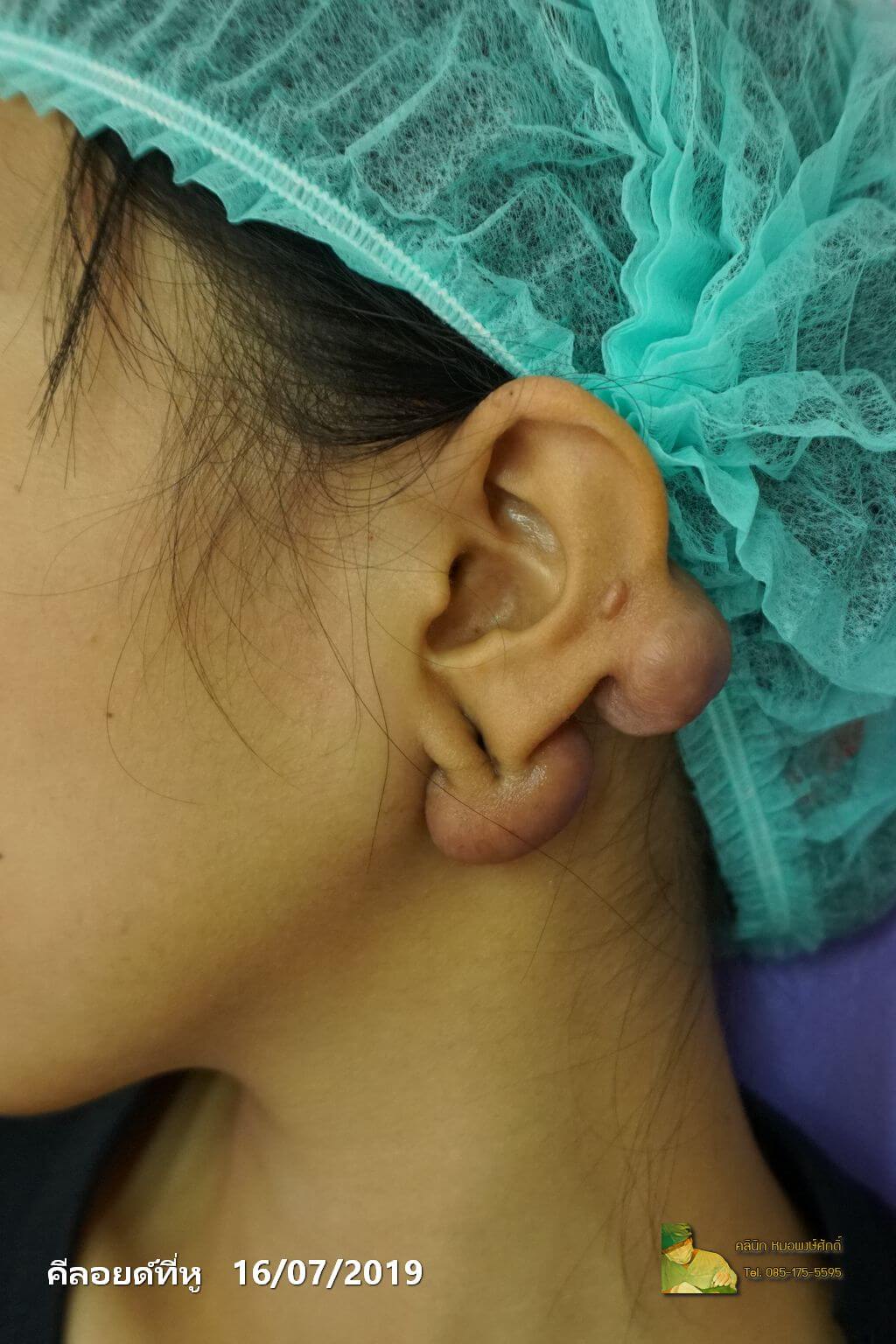
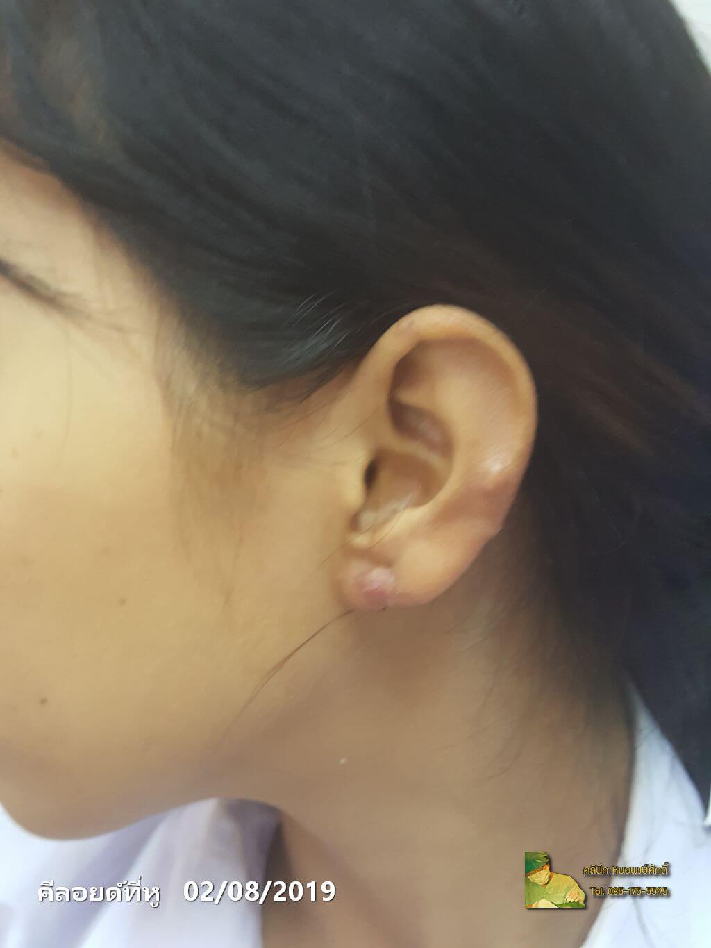
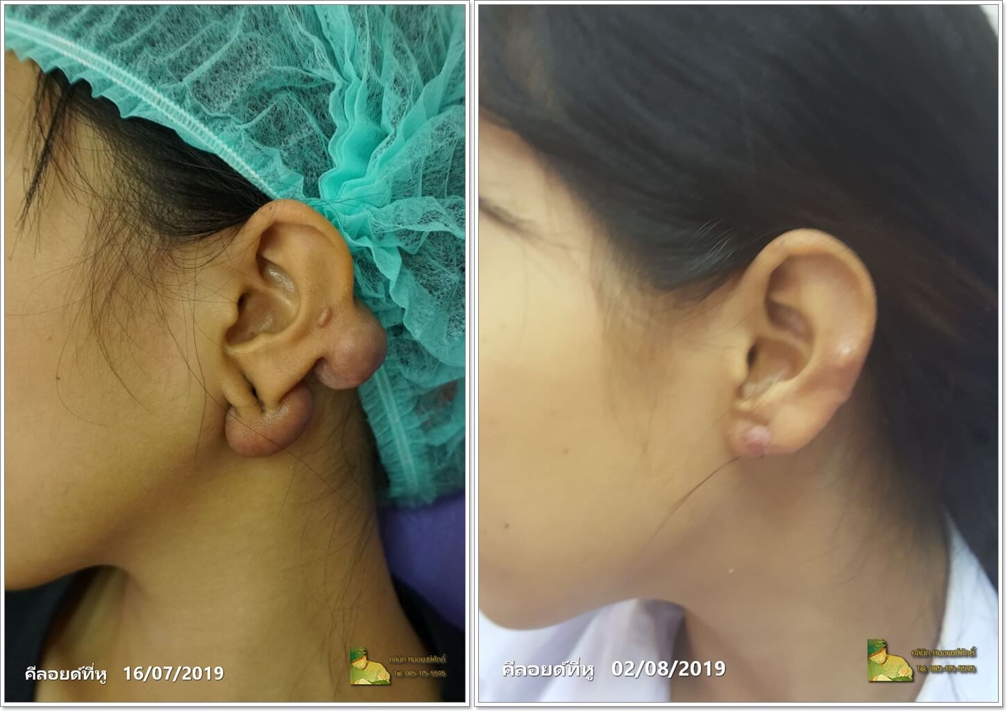
The patient presented with two coexisting keloids on the same ear — a large pedunculated mass projecting from the inferior earlobe and a second nodular lesion within the cavum concha. The lower keloid hung well below the natural lobule margin and was visible from any side view; the conchal lesion distorted the bowl of the ear in profile. Both were removed in a single session under local anaesthesia, using a wedge excision at the lobule to preserve its rounded contour and a circumferential excision at the concha. The follow-up photograph, taken roughly two weeks post-op, shows a normal lobule shape, a flat conchal floor, and only a fine linear scar at each excision site. Performed by Dr. Pongsak at Pongsak Clinic, Hua Hin Road 55.